Tóm tắt Luận án Nghiên cứu bệnh gạo lợn do ấu trùng Cysticercus cellulosae gây ra tại tỉnh tỉnh Sơn La, Điện Biên và biện pháp phòng chống
Trang 1
Trang 2
Trang 3
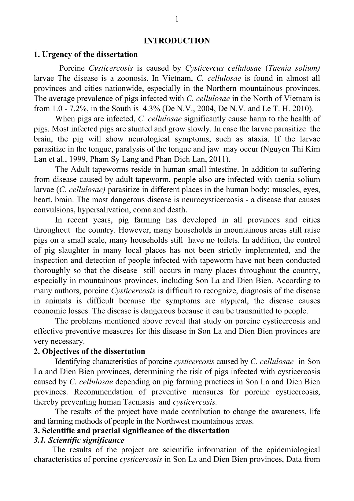
Trang 4
Trang 5
Trang 6
Trang 7
Trang 8
Trang 9
Trang 10
Tải về để xem bản đầy đủ
Bạn đang xem 10 trang mẫu của tài liệu "Tóm tắt Luận án Nghiên cứu bệnh gạo lợn do ấu trùng Cysticercus cellulosae gây ra tại tỉnh tỉnh Sơn La, Điện Biên và biện pháp phòng chống", để tải tài liệu gốc về máy hãy click vào nút Download ở trên.
Tóm tắt nội dung tài liệu: Tóm tắt Luận án Nghiên cứu bệnh gạo lợn do ấu trùng Cysticercus cellulosae gây ra tại tỉnh tỉnh Sơn La, Điện Biên và biện pháp phòng chống
od of larvae in the smoked house at the smoked meat production base as follows: - Lean pork was cut lengthwise into slices of 5 cm thick, 20-25 cm long. The pork was put into pots and 4% salted and mixed for 30 minutes for salt infusion.The pork was placed in a pot and 4% salted, mixed for salt for infusion for about 30 minutes. The pork was placed in a wood burning smokehouse with a temperature of 60 - 65oC, for 24, 48, 72, 96 and 120 hours, ensuring the meat's moisture content of 45%. Number of smoked meat was 15 slices (3 slices were examined per day). Time to examine the vitality of larvae in smoked meat was after 1 day, after 2 days, after 3 days, after 4 days and after 5 days. Method of examination: larvae were examinated whether they still survived or killed by using the above method. 2.4.3.2. Study on treatment regimen of cysticercosis in pigs * Method of evaluating the efficacy of treatment regimen of cysticercosis in pigs a). Designing experiments to assess the efficacy of the treatment regimens for cysticercosis in pigs experimentally infected with cysticercosis 20 pigs were used in the experimental C. cellulosae infection 60 days after infection, Taenia solium ELISA kit from Scimedx/USA was used for serologic test. The results showed that all 20 pigs were seropositive to C. cellulosae antigen. From this, experimental treatment regimens for cysticercosis in pigs were designed. + Group I: 5 pigs were given with praziquantel at, dosage of 20 mg /kg B.W/day for 2 days. Group II: 5 pigs, were given with albendazole, at dosage of 15 mg/kg B.W/day for 3 days,Group III: 5 pigs were given with praziquantel at dosage of 20 mg/kg B.W/day on the first day. Then albendazole was given at dosage of 15 mg\kg\day for the next 2 days. The drug was orally administered. Control group: no drugs were used in 5 pigs. Pigs from 3 experimental groups and control group were dissected on day 11, 12, 13, 14 and 15 after dosing. Evaluating the safety of the cysticercosis treatment regimens: The safety of the cysticercosis treatment regimens were evaluated by monitoring body condition, movement and eating from 15 pigs in 3 experimental groups before and after dosing within 24 hours. b). Evaluating the efficacy of the treatment regimens in infected pigs in the field * Identifying pigs infected with C. cellulosae for treatment (through identifying pigs that were seropositive to antibodies against C. cellulosae. - Using T. solium ELISA kit from Scimedx / USA for serological test of samples from 150 pigs with weak physical condition in Son La and 150 pigs with weak physical condition in Dien Bien. Pigs that were seropositive were considered to get cysticercosis. * Clinical monitoring of seropositive pigs in the field: The pigs showing seropositive by using the ELISA Kit were clinically monitored. Combining the symptoms observed in pigs suffering from natural cysticercosis with the symptoms of pigs experimentally infected with cysticercosis, thereby recognising some relatively typical symptoms of pigs infected with cysticercosis that helped in diagnosis and detection of infected pigs in locations through clinical symptoms of the disease. * Determining the efficacy of the treatment regimens for cysticercosis in pigs in the field: Experimental treatment of pigs that were seropositive showing the most effective regimen selected from test results from infected pigs) . 20 and 30 days after drug administration, serum samples from the treated pigs were retested using Taenia solium ELISA kit. From this, the efficacy of the treatment regimens could be evaluated . 2.4.3.3. Reccomendation of prevention and treatment interventions of cysticercosis in pigs Preventive and control measures of porcine cysticercosis were developed based on epidemiological, pathological and clinical characteristics and experimental results from the disease prevention and treatment studies. 2.4.3.4. Method of developing epidemiological maps Epidemiological map of cysticercosis occuring in pigs in the districts of the two studied provinces was developed using GPS and GIS techniques. 2.4.3.5. Data processing methods The collected data was processed by common mathematical formulas and biological statistical methods of Nguyen Van Thien, 2008), on Excel 2007 and Minitab software 16.0 Chapter 3 RESULTS AND DISCUSSION 3.1. Study on some epidemiological characteristics of porcine cysticercosis caused by Taenia solium larvae (Cysticercus cellulosae) in Son La and Dien Bien provinces 3.1.1. Situation of Cysticercus cellulosae infection in pigs in Son La and Dien Bien provinces 3.1.1.1. The prevalence and intensity of infection with Cysticercus cellulosae in pigs in some districts in Son La and Dien Bien provinces Results of the larval prevalence and intensity were presented in Table 3.1. Table 3.1. The prevalence and intensity of C. cellulosae in some districts of Son La and Dien Bien provinces Location (province, district) Number of dissected pigs (pig) Number of infected pigs (pig) Prevalence of infection (%) Number of pigs nfected with lavae/ intensity of infection (min - max) In skeletal muscles In brain In kidneys In heart * Son La Province 1.040 27 2.59a 27/(1 - 9) 26/(1 - 7) 22/(1 - 4) 19/(1 - 3) Bac Yen District 351 11 3.13 11/(1 - 9) 11/(2 - 7) 10/(1 - 4) 8/(1 - 3) Muong La District 337 9 2.67 9/(1 - 7) 8/(1 - 6) 7/(1 - 3) 5/(1 - 2) Mai Sơn District 352 7 1.99 7/(1 - 9) 7/(1 - 4) 5/(1 - 3) 6/(1 - 2) * Dien Bien Province 1.038 35 3.37a 35/(1 - 10) 32/(1 - 7) 32/(1 - 4) 34/(1 - 4) Muong Ang District 350 10 2.86 10/(1 - 9) 9/(1 - 4) 9/(1 - 3) 9/(1 - 2) Tua Chua District 345 12 3.48 12/(1 - 10) 10/(1 - 5) 10/(1 - 3) 12/(1 - 4) Nam Po District 343 13 3.79 13/(1 - 10) 13/(1 - 7) 13/(1 - 4) 13/(1 - 3) Total 2.078 62 2.98 62/(1 - 10) 58/(1 - 7) 54/(1 - 4) 53/(1 - 4) * Notes: - In Son La province (in Bac Yen district: 11 of 11 infected pigs showed larvae in their muscles and brains, 10 of infected pigs the larvae were found in kidneys, 8 were found in heart. It was Similar to 2 districts of Muong La and Mai Son). - In Dien Bien Province (in Muong Ang district: The larvae were found in 10 of 10 infected pigs with 9 larvae in the brains, kidneys and hearts.The finding was similar to two districts of Tua Chua and Nam Po). - In vertical line, prevalence of infection with the same letters are not significantly different (P> 0.05). Table 3.1 showed that Pigs in Son La and Dien Bien provinces were infected with the larvae at 2.98%. The prevalence of C. cellulosae in pigs in Dien Bien and Son La provinces was not significantly different (P> 0.05).Our study results on the prevalence of C. cellulosae infection in pigs in Son La and Dien Bien provinces were lower than that of Nguyen Quoc Doanh et al. (2002). According to these author, when C. cellulosae was examinated in pigs in Bac Ninh and Bac Kan by using ELISA method, the prevalence of infection in pigs was 9.91%, varying from 6.06 -15.49%. We found that there were differences in practices and techniques of pig rfarrming in various regions, so the prevalence of the larval infection varied. The differences in the above results may be due to different study methods used, the larvae detection method that we performed was dissection of pigs, this method has a very high accuracy, excluding the possibility of cross-reaction that other methods, including ELISA techniques may, encounter. 3.1.1.2. The prevalence of Cysticercus cellulosae infection in pigs depends on age The prevalence of C. cellulosae depending on age of pigs was shown in the column chart in Figure 3.2. Figure 3.2. Figure of prevalence of C. cellulosae infection in pigs depending on age The chart in Figure 3.2 showed that Overall, pigs under 2 months of age C. cellulosae larvae infection were not found . Pigs from 2 - 6 months old were infected with the larvae at a low percentage (1.36%); pigs from 6 - 12 months of age were infected with the larvae with a higher percentage (2.65%), pigs more than 12 months of age had the highest percentage of the larval infection (5.21%). Thus, the prevalence of C. cellulosae infection increased with pig age. This rule is evident in both dissected pigs in Son La and those in Dien Bien province. Gavidia C.M et al. (2013) reported that the prevalence of C. cellulosae increased with the pigs age. Adult pigs were infected more severely than younger ones. Our study results were similar to those of the authors mentioned above. 3.1.1.3. The prevalence of Cysticercus cellulosae in pigs depends on seasons The prevalence of C. cellulosae depending on seasons was shown in Table 3.3 (Main thesis). Table 3.3 showed that the seasons affected the prevalence of C. cellulosae in pigs in Son La and Dien Bien provinces. The percentage of the larval infection, in Summer - Autumn seasons was 3.80%, in Winter - Spring was 2.15%. Thus, the prevalence of C. cellulosae infection in pigs in Summer - Autumn season were higher than those in Winter - Spring season because the Summer - Autumn seasons are hot, humid and rainy creating favourable conditions for tapeworm eggs to exist in the external environment. If people infected with T. solium pass feces containing gravid proglottids into the environment, the gravid proglottids decomposed to release eggs, tapeworm eggs last for quite a long time in the outside. Pigs acquire the infection after ingestion of embryonated eggs or gravid proglottids passed through human faeces 3.1.1.4. The prevalence of Cysticercus cellulosae in pigs depending on the technique of pig farming The results from infection of C. cellulosae prevalence were shown in the chart in Figure 3.4. Figure 3.4. Chart of prevalence of C. cellulosae infection depeding on the technique of pig farming Figure 3.4 showed that: The prevalence of C. cellulosae infection was the highest in free range pigs (5.37%); followed in half free range pigs (1.94%). The lowest prevalence of infection was in pigs raised in confinement (0.66%). This rule was also found in individual pigs dissected separately in each province. We found that in comparison to pigs raised in confinement, free range pigs and half free range pigs have risk to expose to tapeworm eggs passing through human faeces causing higher prevalence of C. Cellulosae infection 3.1.1.5. Prevalence of Cysticercus cellulosae in crossbred pigs and local pigs The results from prevalence of C. Cellulosae infection were shown in Table 3.5. Table 3.5 showed that in the two provinces as a whole, the prevalence of the larval infection in local pigs was higher than that of cross bred pigs (3.84% compared to 1.98% respectively). In particular, In Son La, the prevalence of C. cellulosae infection in local pigs was 3.19%, in cross bred pigs was 1.89%. In Dien Bien, the prevalence of C. cellulosae infection in local pigs was 4.49% in cross bred pigs was 2.08%. Thus, according to the results mentioned above, the prevalence of infection between local and crossbred pigs varied. However, field surveys showed that local pigs mostly were free range and halffree range while crossbred pigs were mostly raised in confinement. Therefore, the larval infection in the local pig was higher than that of crossbred pigs due to technique of pig farming. Therefore in order to determine whether the prevalence of larval infection depends on pig breeds, we continued to monitor the prevalence of larval infection in both the free range local pigs and crossbred pigs. The results were shown in the chart in Figure 3.6. Figure 3.6. Prevalence of C. cellulosae infection in the free range local pigs and crossbred pigs The chart in Figure 3.6. Showed that in the two provinces, the prevalence of larval infection in cross bred pigs and local pigs was similar when they were raised free range (5.43% compared to 5.33% respectively). Borkataki S et al. (2011) reported that the prevalence of lavae infection in cross-bred pigs and local pigs was 12.53% and 7.49% respectively. Our study finding was different from those of the authors mentioned.above In our opinion, when pigs are raised with the same method, the breed factor almost does not affect the prevalence of C. Cellulosae infection. 3.1.2. Actual situation of Taenia solium infection in humans in some districts of Son La and Dien Bien provinces 3.1.2.1. Investigation results from the current status of livestock production practices and people's living activities in some districts of Son La and Dien Bien provinces Investigation results from the current status of livestock production practices and people's living activities related to prevalence of cysticercosis in pigs and T. solium infection in humans were shown in Table 3.8. Table 3.8 showed: that in Son La province, households raising pigs free range acconted for 71.34%. households using toilets acconted for 64.53%, but many of them whose toilets were very sparse and flimsy. In Dien Bien province: households raising pigs free range accounted for 73.73%. housseholds using toilets accounted 58.67%. Through the investigation results we found that people's living activities and the livestock production practices were still insufficient, facilitating spreading parasitic diseases in general, and porcine cysticercosis and human Taeniasis (T. solium infection) in in particular. 3.1.2.2. Investigation results from the current status of people's eating habits in some districts of Son La and Dien Bien provinces Investigation results from people's eating habits related to the prevalence of C. cellulosae infection in pigs and T. solium infection in humans were shown in Table 3.9. Table 3.9 showed: that In Son La province people eating undercooked meat, rare meat, and smoked meat accounted for a high proportion 44.27%; 69.07% respectively. In Dien Bien province, these proportions were 41.2% and 81.07% respectively. From the above results, we realized that local authorities should propagate ethnic minorities not to eat rare meat or undercooked meat so that they can change their eating habits available from the ancient time; When smoked meat used, it must be cooked to decrease the prevalence of cestode infection in humans, simultaneously prevention of cestode infection in humans, which is an active measure for prevention of porcine cysticercosis; 3.1.2.3. The prevalence of tapeworm infection among investigated people in some districts of Son La and Dien Bien provinces The prevalence of T. solium infection in humans among the investigated people in the two provinces was shown in Table 3.10. Table 3.10 showed that the prevalence of tapeworm infection among investigated people in 3 districts of Son La province was 3.07%. The prevalence of T. solium infection among investigated people in 3 districts of Dien Bien province was 3.86%. Through the investigation, we found that Bac Yen district of Son La province and Nam Po district of Dien Bien province are remote areas of these two provinces, economic conditions, difficult transportation, and knowledge of human T. solium and cysticercosis in pigs are very limited, preventive measure of the disease is not implemented, so the proportion of people infected with cestode was higher than that of the remaining districts. According to the Ministry of Health (2019), the prevalence of cestode infection in humans in the midland and mountainous areas was 2 - 6%. Thus, the investigation results on the prevalence of cestode infection in humans in Son La and Dien Bien provinces were also within the rate limits announced to the public by the Ministry of Health. 3.1.5. Determining the correlation between Taenia solium infection and the prevalence of Cysticercus cellulosae in pigs in Son La and Dien Bien The correlation between the prevalence of T. solium infection in humans and the one of C. cellulosae larval infection in pigs in Son La and Dien Bien was shown in Table 3.16 and charts in Figure 3.11a and 3.11b. * In Son La Province: The correlation between prevalence of T. solium infection in humans and the prevalence of C. cellulosae infection in pigs was positively correlated. The correlation coefficient R = 0.929 proves that the correlation is very tight. * In Dien Bien Province: The correlation between the prevalence of T. solium infection in humans and the prevalence of C. cellulosae in pigs was also positively correlated. The correlation coefficient R = 0.74 indicated that the correlation was relatively tight. 3.2. Study on cysticercosis in experimental infection of pigs 3.2.1. Examination of Taenia solium to cause cysticercosis in pigs * Results of comparing the similarity of nucleotide sequence of cestode samples and standard samples The CO1 gene sequences of 6 cestode samples were with 396 bp in length. The comparison results showed that the sequences were completely similar to each other, which were shown in Figure 3.12. Compared to the sequences on the Genbank (Basic Local Alinment Search Tool) by the BLAST program, these sequences had a high similarity (100%) with those of T. solium species, which were shown in Table 3.17. According to BLAST results, the CO1 gene sequences of the studied samples had 100% resemblance of those of T. solium species. Genetic gap analysis showed that the CO1 gene sequences of the six T. solium samples collected from Son La and Dien Bien (Vietnam) showed high similarity (100%) with the sequences of this species in China, Indonesia and India, which were shown in Table 3.18. On the phylogenetic tree (Figure 3.13), the CO1 gene sequence of T. solium species in Vietnam was in the same branch as the CO1 gene sequence of T. solium species in China, Indonesia and India. Figure 3.13. A phylogenetic tree is constructed from the CO1 gene sequence using the Maximum Likekliwood method 3.2.2. The results of experimentally Infected cysticercosis in pigs were shown in Table 3.19. Table 3.19 showed that Pigs in experimental groups I and II, 90 - 120 days after infection, all of the larvae were fully developed at the parasitic site. Number of cestode eggs infecting group I and group II was different so number of larvae at parasitic sites was also different. The pigs in group II had more larvae at parasitic sites than pigs in group I with manifestation of all symptoms.mentioned previously. According to Pham Van Khue and Phan Luc (1996), Pham Van Than (2009), Nguyen Thi Kim Lan (2012), the time period for C. cellulosae to fully develop in pigs was 60 days. Thus, our results from the larvae development time period was longer than published papers of these authors. 3.2.2.1. Clinical signs in pigs with experimental cystierticosis Table 3.20. Clinical signs in pigs with experimental Cysticercosis Experimental group Number Of pigs (pig) Number Of pigs manifesting clinical signs (pig) percentage of pigs manifesting clinical signs (%) Cinical signs Major clinical signs Number Of pigs (pig) Percentage (%) Expermentally Cysticercosis ìnfected group I 7 7 100 Fever 4 57.14 Cough 5 71.43 Diarrhea 4 57.14 Sore eyes and eye dischage 5 71.43 Anorexia, slow moving 4 57.14 Convulsion, salivasion 2 28.57 Skinny, Pale mucosa, ruffled hair coat 7 100 Erectile neck hairs trembling, grinding teeth 7 100 Difficulty walking with a limp 3 42.85 Experimentally Cysticercosis ìnfected group II 7 7 100 Fever 7 100 Dyspnea, cough 6 85.71 Diarrhea 5 73.43 Sore eyes with rheum 7 100 Anorexia 6 85.71 Convulsion,salivasion 4 57,14 Skinny, Pale mucosa, ruffled hair coat 7 100 Erectile neck hairs trembling, grinding teeth 7 100 Difficulty walking with a limp 5 71.43 Control group 7 0 0 0 0 0.00 The results in Table 3.20: showed that In experimental group I and II:1 - 10 days after infection, pigs showed clinical signs such as fever, cough, skinny, pale mucous membranes, ruffled hair coats, diarrhea, eye pain with discharge, convulsions, hypersalivasion, erected hair on the back of the neck, trembling, g
File đính kèm:
 tom_tat_luan_an_nghien_cuu_benh_gao_lon_do_au_trung_cysticer.doc
 TRANG THONG TIN LUAN AN TS DO THI LAN PHUONG.doc
 TRICH YEU LUAN AN DO THI LAN PHUONG.dot

